UNIT 1 – DIGESTION AND
ABSORPTION OF FOOD
SL – STRUCTURE AND
FUNCTION OF DIGESTIVE GLANDS[i]
NERVOUS SYSTEM
The nervous system is divided into
1.
Central nervous system (CNS)
2.
Peripheral nervous system (PNS)
2.1. Somatic nervous system (SoNS)
2.2. Autonomic
nervous system (ANS)
2.2.1.
Sympathetic nervous system (SNS)
2.2.2.
Parasympathetic nervous system (PsNS)
1.
Central nervous system (CNS) - it comprises of the brain and spinal cord –
also called as the body’s master control unit
2.
Peripheral nervous system (PNS) – it includes
all the nerves arising out and going to the central nervous system – also
called the body’s link to the outside world
2.1. Somatic nervous system (SoNS): cerebrospinal-
it is a part of the peripheral nervous system, is associated with the voluntary
control of body movements via skeletal muscles. The somatic nervous system
consists of (afferent nerves) sensory nerves and (efferent nerves) motor
nerves. The somatic nervous system controls all voluntary muscular systems
within the body, and the process of voluntary (somatic) reflex arcs.[1]
2.2. Autonomic
nervous system (ANS): visceral
2.2.1.
Sympathetic nervous system (SNS) – the action of
most of the organs is accelerated by the SNS
2.2.2.
Parasympathetic nervous system (PsNS) – the
activity of most of the organs are inhibited by PsNS
It must by however
noted that, all the nervous activities are always controlled by the central
nervous system; the peripheral nervous system are merely carriers of the
information.
Only a small part of the body activities are under the
willful (coluntary) control of the body. The centers for the control of
voluntary activities are present in the thalamus and cerebral cortex of the
brain. These are therefore called as the conscious areas of the brain.
Most of the organs of the body are controlled by an automatic feedback circuit
in which no conscious thinking is required. Such activities are termed as
involuntary and are controlled by autonomic nervous system. The centers
for involuntary actions are present in the medulla oblongata, pons
and midbrain.
The SNS regulates actions that require quick action and PsNS
calms down the actions of SNS.
The PsNS regulates the actions that do not require quick
responsiveness.
1.
Motor functions
2.
Sensory functions
DIGESTIVE SYSTEM
Digestion is the mechanical and chemical breakdown of foods
into forms that the cell membranes can absorb.
Mechanical digestion breaks large pieces into smaller ones
without altering their chemical composition
Chemical digestion breaks food into simpler chemicals.
The digestive system carries out ingestion, propulsion,
digestion, absorption and defecation.
The digestive system consists of the alimentary canal,
extending from the mouth to the anus, and several accessory organs, which
release secretions into the canal.
The alimentary canals includes the mouth, pharynx,
oesophagus, stomach, small intestine, large intestine, and anal canal.
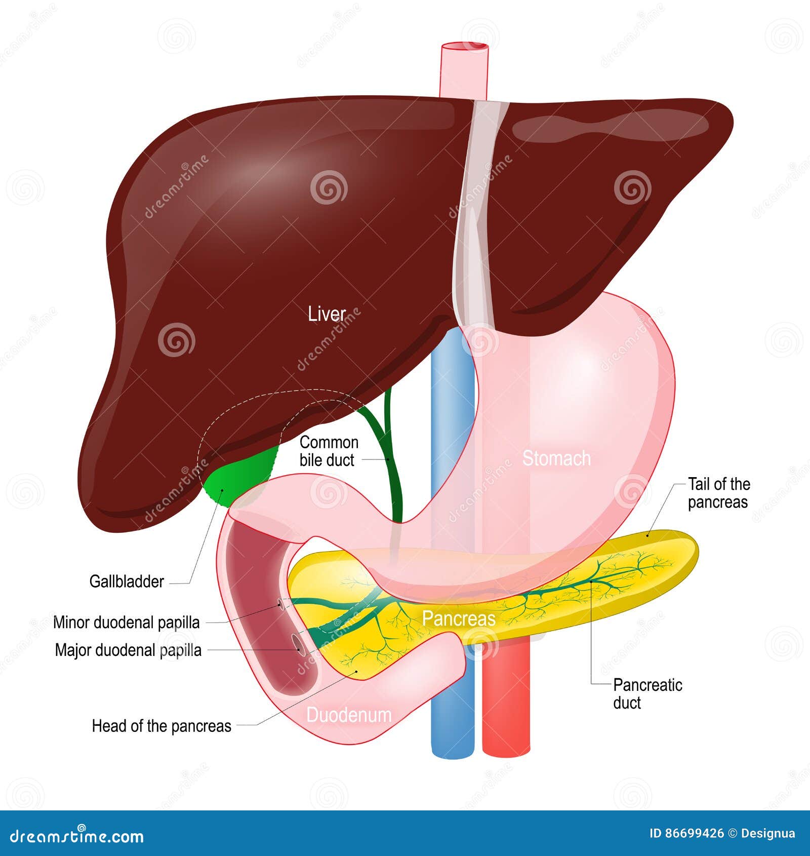
The accessory organs include the salivary glands, liver,
gall bladder and pancreas.
The digestive system originates from the inner layer
(endoderm) of the embryo, which folds to form the tube of the alimentary canal.
The accessory organs develop as the buds from the tube.
DIGESTIVE SYSTEM
The digestive system consists of the
alimentary canal, extending from the mouth to the anus, and several accessory
organs, which release secretions into the canal. The alimentary canal includes
the mouth, pharynx, esophagus, stomach, small intestine, large intestine and
anal canal. The accessory organs include the salivary glands, liver, gall
bladder and pancreas. The digestive system originates from the inner layer
(endoderm) of embryo, which folds to form tube of alimentary canal. The accessory
organs develop as buds from the tube.
The mouth is followed by a muscular tube
(Pharynx). The pharynx leads to a spacious chamber – stomach, through a narrow
tube – oesophagus. The passage from the oesophagus to the stomach is guarded by
a valve known as cardiac
spinchter. The stomach is followed
by small intestine which can be divided into three parts – duodenum, jejunum,
ileum.
The distal end of the stomach opens through pyloric spinchter into the duodenum. The middle part is
jejunum and the last part is ileum.
The small intestine (ileum) opens into the
large intestine (colon) through ileo-colic
valve.
A vermiform appendix is present at the site
of ileo-colic valve which is vestigial in human beings.
The colon leads into the last part of
intestine – rectum. The rectum opens outside the body, the rectal opening is
guarded by anal spinchter.


GENERAL PLAN OF THE ALIMENTARY CANAL
The alimentary canal is a muscular tube about 8 meters long
that passess through the thoracic and abdominopelvic cavities. The structure of
its walls, function and innervations are similar throughout its length with
slight modifications at places.
Structure of the wall
The wall of alimentary canal consists of four distinct
layers that are developed to different degrees from region to region. The four
distinct layers persists throughout the alimentary canal, but certain regions
are specialized for particular functions. Beginning with innermost tissues, the
layers are as follows.
1.
Mucosa
layer
This layer is formed of surface epithelium,
underlying connective tissue (lamina
propria), and a small amount of smooth muscles (muscularis mucosa). In some regions the mucosa is folded with tiny
projections towards lumen. This increases the absorptive surface area.
The mucosa has tubular invaginations, which
are lined by cells that secrete mucus and digestive enzymes.
2.
Submucosa
layer
Next to the mucosa layer is the submucosa
layer. It contains considerable loose connective tissue as well as glands,
blood vessels, lymphatic vessels, and nerves. Its blood vessels nourish the
surrounding tissues and carry away absorbed materials.
3.
Muscular
layer
This layer, which provides movement of the
tube, consists of two layers of smooth muscle tissues. The outer is the layer
of longitudinal muscle. The inner is the layer of circular muscle. The
contraction of circular muscle fibers reduces the diameter of lumen of the
alimentary canal; the contraction of longitudinal muscle layers shortens the
length of the tube.
4.
Serosa
The serosa layer is the outer covering of
the tube; it is composed of the visceral
peritoneum, which is formed of epithelium on the outside and connective
tissue beneath. The cells of serosa protect the underlying tissue and secrete
serous fluid, which moistens and lubricates the tubes outer surface so that the
organs (which are lined by parietal peritoneum) slide freely inside the body
cavity and against one another.
5.
Gut associated lymphoid tissues (GALT) –
Peyer’s patches
Innervation [6]of
the tube
Branches of the sympathetic and parasympathetic divisions of
autonomic nervous system extensively innervates the alimentary canal. These
nerve fibers, mainly associated with tube’s muscular layer, maintain muscle and
regulate the strength, rate and velocity of muscular contractions.
Many of the postganglionic
fibers are organized into a nerve plexus [7]within
the wall of the canal.
The submucosal
(Meissner’s plexus) plexus is important in controlling secretions of
gastrointestinal tract.
The myenteric
plexus of the muscular layer controls the gastrointestinal motility
The nerve plexus of the gastrointestinal tract are so
extensive, that it is some times said to have a ‘second brain’.
** a great
advance in our knowledge of gastric digestion, particularly in man, was made
through the observations of Beaumont on his patient, Alexis St.
Martin who in 1822, following a gunshot wound was left with an opening from
the stomach through the abdominal wall to the exterior. Through this fistula,
Beaumont found it possible to follow the course of gastric digestion of
different food under varying conditions of health and obtained pure gastric
juice for digestion experiments outside body
Movements of the tube
The motor functions
of the alimentary canal are of two basic types – mixing movements and propelling
movements
Mixing occurs when
smooth muscles in particular segments of tube contract and relax rhythmically.
For example – when the stomach is full, waves of the muscular contractions move
along its wall from one end to the other. These waves occur every twenty
seconds of so. They mix the digestive juices secreted by the mucosa with food.
Propelling movements
include a wavelike motion called – peristalsis.
During peristalsis a ring of
contraction appears in the wall of the tube. At the same time the the muscular
wall just ahead of the ring relaxes – a phenomena called as receptive
relaxation.
Law of gut: when a segment of the intestinal tract is
excited by distention and thereby initiates peristalsis (peristaltic reflex),
the contractile ring causing the peristalsis normally begins on the orad [8]side of
the distended segment and moves towards the distended segment, pushing the intestinal
contents in the anal direction for 5 – 10 centimeters before dying out. At the
same time the gut relaxes several centimeters downstream toward the anus
(receptive relaxation), thus allowing the food to be propelled easily towards
the anus.
This complex
movement occurs only in the presence of the myenteric plexus. Therefore the
movement is called the myenteric reflex or peristaltic reflex.
The peristaltic
reflex and the movement of peristalsis towards anus is called the “law of gut”
[1]
There are two types of reflex
arcs; the autonomic reflex arc – affecting inner organs and the somatic
(voluntary) reflex arc – affecting skeletal muscles.
[6]
Supply nerves to; to put the
nerves into
[7]
network
[8]
Towards the mouth; oral
direction
[i]
Guyton, Hall, Textbook of
Medical Physiology, 11th Edition, Elsevier
Shier
D, Butler J, Lewis R – Hole’s Human anatomy and Physiology, 11th
Edition
Agarwal,
Srivastava, Kumar – Animal Physiology and Biochemistry, 5th Edition,
2013, S. Chand & Company Ltd
Verma,
Tyagi, Agarwal – Animal Physiology, 2015, S. Chand & Company Ltd
Asim
Kumar Datta – Functional Histology, 1st Edition, Current Books
International

