Monday, June 14, 2021
Sunday, March 17, 2019
ORDER HEMIPTERA
The hemiptera are a large frequently
encountered order, members of which are extremely diverse in size shape and
colour. They include insects commonly known as bugs, leafhoppers, cicadas,
aphids, lerps and scale insects. The most distinctive feature of hemipterans is
their sharply pointed, tube-like mouthparts (proboscis or rostrum) that are
used for piercing or sucking. Hemipterans usually have two paier of wings,
however some groups may be wingless and others have only forewings. They feed
on juices of plants or animals.
The Order Hemiptera is divided into
four Suborders
1.
Heteroptera (true
bugs)
The term heteroptera, derived from
greek, hetero-different; ptera-wings; The forewings are hardened at the base,
membranous at the tips, and sitting flat over abdomen, hiding the membranous
hind wings; the head and proboscis can flex forward and some predatory species
have raptorial forelegs.
Wings lie flat on the back at rest,
forming an ‘X’
Figure 1: Heteroptera: Leptocorisa
2.
Auchenorrhyncha (cicadas, spittlebugs, leafhoppers, planthoppers and tree hoppers)
They have forewings uniform in
texture and held like a tent over the abdomen; the head and proboscis are
directed down and back and many have hind legs adapted for jumping.
Figure 2: Auchenorrhyncha: Nilaparvata lugens
3.
Sternorrhyncha (psyllids, whiteflies, aphids, mealy bugs)
They are usually small, soft bodied
and generally wingless; the head and proboscis are directed down and back, and
in some the legs are vestigial or absent. Many species cover themselves with
wax to prevent their soft bodies from dessicating.
Figure 3: Sternorrhyncha: Cereal aphid
4.
Coleorrhyncha (moss
bugs)
They are small, rarely seen, group of
flattened, mostly flightless bugs that are found amongst mosses and liverworts.
Blood Sucking Bugs (Order Hemiptera; Suborder Heteroptera)
BLOOD SUCKING BUGS
FAMILY CIMICIDAE (BED BUGS)
The Cimicidae form a well-defined family of blood sucking bugs. They are
oval flattened insects without functional wings, although the forewings remain present
as two small pads on the dorsal surface of the thorax.
There are 91 recognized species of Cimicidae.
·
Most
are associated with birds and/or bats but two species Cimex lectularius
and Cimex hemipterus are the familiar bedbugs commonly associated with
man.
·
Both
species lay their eggs in cracks and crevices of houses and outbuildings.
·
Each
female bedbug lays about 200 eggs, which hatch about after 10 days at 20 degree
Celsius.
·
The
nymphs and adults usually feed at night when their hosts are sleeping, although
they will feed during the day if conditions are favourable.
·
Feeding
behaviour, and hence development, is critically dependent on temperature and
humidity.
·
Bedbugs
do not feed at temperatures less than 13 degree Celsius.
·
Experimental
evidences show that bedbugs can be infected with a range of human parasites and
pathogens, including hepatitis-B, HIV and Trypanosoma cruzi.
·
Bedbugs
will feed daily if given the opportunity and their high numbers can contribute
to chronic iron-deficiency anaemia, especially in infants.
FAMILY
POLYCTENIDAE (BAT BUGS)
The family
polyctenidae comprises 32 species grouped in five genera. They are small ectoparasites
of bats, with no known medical importance. They lack eyes and ocelli and are
always flightless with forewings reduced to small flaps. The bugs are
parthenogenetic. Bat bug Hesperoctenes.
FAMILY
REDUVIIDAE & AND SUBFAMILY TRIATOMINAE (KISSING BUGS)
Most reduviidae
are predators on insects and other invertebrates. They are predominantly
tropical, occupying a very wide range of terrestrial habitats and displaying a
variety of hunting strategies and pre preferences. Over 6000 species are known,
which are grouped into 23 subfamilies. In many predatory reduviids, the fore
legs are adapted to hold prey. Often the fore legs (sometimes mid legs) are
strongly raptorial, equipped with spines, adhesive organs and/or glands
secreting a glue-like substance.
Subfamily
Triatominae (Kissing bugs)
There are
118 species of Triatominae recognized on the basis of morphological characters.
They range from 5mm to 45 mm in length.
All species
of triatominae are obligate bloodsuckers and over half have been shown
naturally or experimentally to be susceptible to infection with Trypanosoma
cruzi.
Epidemiologically
only about a dozen species have become sufficiently closely associated with man
to represent a public health problem; of these the most important vector
species are Triatoma infestans, Panstrongylus megistus, Rhodnius prolixus,
Triatoma brasiliensis and Triatoma dimidiata.
Other species
are mainly associated with nest-building birds and small mammals, and occasionally
reptiles.
References
·
Medical Insects and Arachnids Edited by
Richard P. Lane and Roger W. Crosskey. Published in 1993 by Chapman & Hall
ISBN 0 412 400006
Tuesday, January 8, 2019
DIGESTIVE SYSTEM: INTRODUCTION AND GENERAL PLAN OF ALIMENTARY CANAL
UNIT 1 – DIGESTION AND
ABSORPTION OF FOOD
SL – STRUCTURE AND
FUNCTION OF DIGESTIVE GLANDS[i]
NERVOUS SYSTEM
The nervous system is divided into
1.
Central nervous system (CNS)
2.
Peripheral nervous system (PNS)
2.1. Somatic nervous system (SoNS)
2.2. Autonomic
nervous system (ANS)
2.2.1.
Sympathetic nervous system (SNS)
2.2.2.
Parasympathetic nervous system (PsNS)
1.
Central nervous system (CNS) - it comprises of the brain and spinal cord –
also called as the body’s master control unit
2.
Peripheral nervous system (PNS) – it includes
all the nerves arising out and going to the central nervous system – also
called the body’s link to the outside world
2.1. Somatic nervous system (SoNS): cerebrospinal-
it is a part of the peripheral nervous system, is associated with the voluntary
control of body movements via skeletal muscles. The somatic nervous system
consists of (afferent nerves) sensory nerves and (efferent nerves) motor
nerves. The somatic nervous system controls all voluntary muscular systems
within the body, and the process of voluntary (somatic) reflex arcs.[1]
2.2. Autonomic
nervous system (ANS): visceral
2.2.1.
Sympathetic nervous system (SNS) – the action of
most of the organs is accelerated by the SNS
2.2.2.
Parasympathetic nervous system (PsNS) – the
activity of most of the organs are inhibited by PsNS
It must by however
noted that, all the nervous activities are always controlled by the central
nervous system; the peripheral nervous system are merely carriers of the
information.
Only a small part of the body activities are under the
willful (coluntary) control of the body. The centers for the control of
voluntary activities are present in the thalamus and cerebral cortex of the
brain. These are therefore called as the conscious areas of the brain.
Most of the organs of the body are controlled by an automatic feedback circuit
in which no conscious thinking is required. Such activities are termed as
involuntary and are controlled by autonomic nervous system. The centers
for involuntary actions are present in the medulla oblongata, pons
and midbrain.
The SNS regulates actions that require quick action and PsNS
calms down the actions of SNS.
The PsNS regulates the actions that do not require quick
responsiveness.
1.
Motor functions
2.
Sensory functions
DIGESTIVE SYSTEM
Digestion is the mechanical and chemical breakdown of foods
into forms that the cell membranes can absorb.
Mechanical digestion breaks large pieces into smaller ones
without altering their chemical composition
Chemical digestion breaks food into simpler chemicals.
The digestive system carries out ingestion, propulsion,
digestion, absorption and defecation.
The digestive system consists of the alimentary canal,
extending from the mouth to the anus, and several accessory organs, which
release secretions into the canal.
The alimentary canals includes the mouth, pharynx,
oesophagus, stomach, small intestine, large intestine, and anal canal.
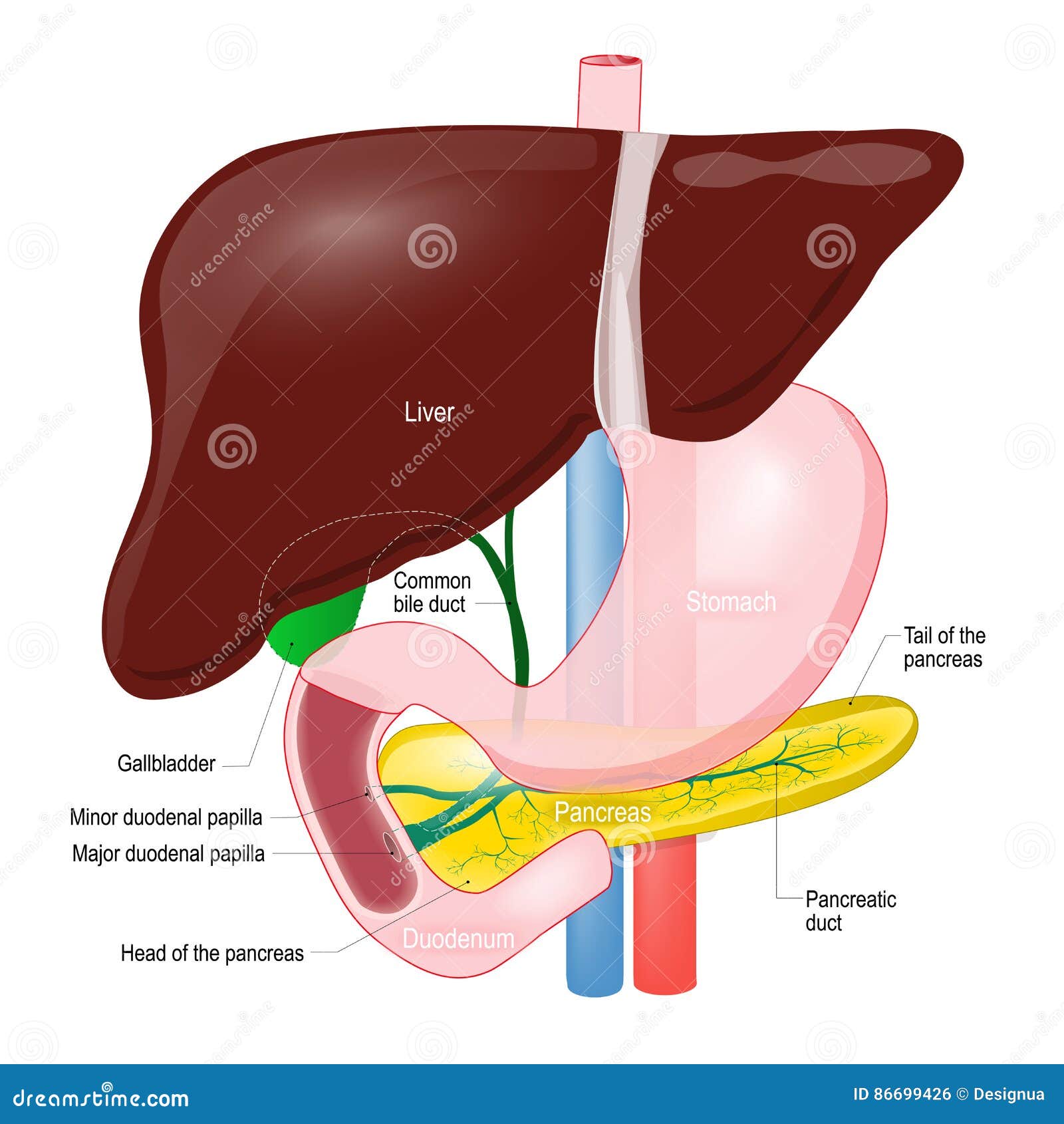
The accessory organs include the salivary glands, liver,
gall bladder and pancreas.
The digestive system originates from the inner layer
(endoderm) of the embryo, which folds to form the tube of the alimentary canal.
The accessory organs develop as the buds from the tube.
DIGESTIVE SYSTEM
The digestive system consists of the
alimentary canal, extending from the mouth to the anus, and several accessory
organs, which release secretions into the canal. The alimentary canal includes
the mouth, pharynx, esophagus, stomach, small intestine, large intestine and
anal canal. The accessory organs include the salivary glands, liver, gall
bladder and pancreas. The digestive system originates from the inner layer
(endoderm) of embryo, which folds to form tube of alimentary canal. The accessory
organs develop as buds from the tube.
The mouth is followed by a muscular tube
(Pharynx). The pharynx leads to a spacious chamber – stomach, through a narrow
tube – oesophagus. The passage from the oesophagus to the stomach is guarded by
a valve known as cardiac
spinchter. The stomach is followed
by small intestine which can be divided into three parts – duodenum, jejunum,
ileum.
The distal end of the stomach opens through pyloric spinchter into the duodenum. The middle part is
jejunum and the last part is ileum.
The small intestine (ileum) opens into the
large intestine (colon) through ileo-colic
valve.
A vermiform appendix is present at the site
of ileo-colic valve which is vestigial in human beings.
The colon leads into the last part of
intestine – rectum. The rectum opens outside the body, the rectal opening is
guarded by anal spinchter.


GENERAL PLAN OF THE ALIMENTARY CANAL
The alimentary canal is a muscular tube about 8 meters long
that passess through the thoracic and abdominopelvic cavities. The structure of
its walls, function and innervations are similar throughout its length with
slight modifications at places.
Structure of the wall
The wall of alimentary canal consists of four distinct
layers that are developed to different degrees from region to region. The four
distinct layers persists throughout the alimentary canal, but certain regions
are specialized for particular functions. Beginning with innermost tissues, the
layers are as follows.
1.
Mucosa
layer
This layer is formed of surface epithelium,
underlying connective tissue (lamina
propria), and a small amount of smooth muscles (muscularis mucosa). In some regions the mucosa is folded with tiny
projections towards lumen. This increases the absorptive surface area.
The mucosa has tubular invaginations, which
are lined by cells that secrete mucus and digestive enzymes.
2.
Submucosa
layer
Next to the mucosa layer is the submucosa
layer. It contains considerable loose connective tissue as well as glands,
blood vessels, lymphatic vessels, and nerves. Its blood vessels nourish the
surrounding tissues and carry away absorbed materials.
3.
Muscular
layer
This layer, which provides movement of the
tube, consists of two layers of smooth muscle tissues. The outer is the layer
of longitudinal muscle. The inner is the layer of circular muscle. The
contraction of circular muscle fibers reduces the diameter of lumen of the
alimentary canal; the contraction of longitudinal muscle layers shortens the
length of the tube.
4.
Serosa
The serosa layer is the outer covering of
the tube; it is composed of the visceral
peritoneum, which is formed of epithelium on the outside and connective
tissue beneath. The cells of serosa protect the underlying tissue and secrete
serous fluid, which moistens and lubricates the tubes outer surface so that the
organs (which are lined by parietal peritoneum) slide freely inside the body
cavity and against one another.
5.
Gut associated lymphoid tissues (GALT) –
Peyer’s patches
Innervation [6]of
the tube
Branches of the sympathetic and parasympathetic divisions of
autonomic nervous system extensively innervates the alimentary canal. These
nerve fibers, mainly associated with tube’s muscular layer, maintain muscle and
regulate the strength, rate and velocity of muscular contractions.
Many of the postganglionic
fibers are organized into a nerve plexus [7]within
the wall of the canal.
The submucosal
(Meissner’s plexus) plexus is important in controlling secretions of
gastrointestinal tract.
The myenteric
plexus of the muscular layer controls the gastrointestinal motility
The nerve plexus of the gastrointestinal tract are so
extensive, that it is some times said to have a ‘second brain’.
** a great
advance in our knowledge of gastric digestion, particularly in man, was made
through the observations of Beaumont on his patient, Alexis St.
Martin who in 1822, following a gunshot wound was left with an opening from
the stomach through the abdominal wall to the exterior. Through this fistula,
Beaumont found it possible to follow the course of gastric digestion of
different food under varying conditions of health and obtained pure gastric
juice for digestion experiments outside body
Movements of the tube
The motor functions
of the alimentary canal are of two basic types – mixing movements and propelling
movements
Mixing occurs when
smooth muscles in particular segments of tube contract and relax rhythmically.
For example – when the stomach is full, waves of the muscular contractions move
along its wall from one end to the other. These waves occur every twenty
seconds of so. They mix the digestive juices secreted by the mucosa with food.
Propelling movements
include a wavelike motion called – peristalsis.
During peristalsis a ring of
contraction appears in the wall of the tube. At the same time the the muscular
wall just ahead of the ring relaxes – a phenomena called as receptive
relaxation.
Law of gut: when a segment of the intestinal tract is
excited by distention and thereby initiates peristalsis (peristaltic reflex),
the contractile ring causing the peristalsis normally begins on the orad [8]side of
the distended segment and moves towards the distended segment, pushing the intestinal
contents in the anal direction for 5 – 10 centimeters before dying out. At the
same time the gut relaxes several centimeters downstream toward the anus
(receptive relaxation), thus allowing the food to be propelled easily towards
the anus.
This complex
movement occurs only in the presence of the myenteric plexus. Therefore the
movement is called the myenteric reflex or peristaltic reflex.
The peristaltic
reflex and the movement of peristalsis towards anus is called the “law of gut”
[1]
There are two types of reflex
arcs; the autonomic reflex arc – affecting inner organs and the somatic
(voluntary) reflex arc – affecting skeletal muscles.
[6]
Supply nerves to; to put the
nerves into
[7]
network
[8]
Towards the mouth; oral
direction
[i]
Guyton, Hall, Textbook of
Medical Physiology, 11th Edition, Elsevier
Shier
D, Butler J, Lewis R – Hole’s Human anatomy and Physiology, 11th
Edition
Agarwal,
Srivastava, Kumar – Animal Physiology and Biochemistry, 5th Edition,
2013, S. Chand & Company Ltd
Verma,
Tyagi, Agarwal – Animal Physiology, 2015, S. Chand & Company Ltd
Asim
Kumar Datta – Functional Histology, 1st Edition, Current Books
International
Tuesday, September 11, 2018
ORIGIN & EVOLUTION OF REPTILES
ORIGIN
& EVOLUTION OF REPTILES
Reptiles
evolved from amphibians of Carboniferous period[1], which depended on water
bodies for laying eggs and development of larval stages and hence could not
exploit arid/terristrial habitats far away from water bodies.
They developed
a large yolk-laden [2]shelled egg that could be laid
on land and in which an amniotic[3] sac contained fluid in which
embryo could develop to an advanced stage, capable of fending for itself when
hatched. The following anatomical changes transformed the ancestral amphibians
into land adapted reptiles:
- Body developed a covering of epidermal scales to prevent loss of body moisture, and skin became cornified and devoid of glands.
- Skull became monocondylic[4] for better movement and flexibility. Atlas and axis vertebrae together permitted skull movement in all directions.
- Limb bones and girdles became stronger but limbs were attached on the sides of body, and belly touched the ground during creeping mode of locomotion.
- Sacral region involved two strong and fused vertebrae to support the body weight on hind legs.
- Pentadactyle limbs developed claws that helped in climbing on rocks and trees.
- Respiration through lungs became more efficient to use the oxygen available in the air.
- As a water conservation strategy, metanephric kidneys excreted uric acid which did not require water for excretion.
- Reptiles continued to be ectothermal since ventricle was not completely partitioned[5] by a septum and blood mixed in heart.
- Internal fertilization evolved as a large cleioid[6] shelled egg was laid on land.
- Embryonic membranes amnion, allantois and yolk sac evolved to enable embryonic development in arid conditions.
ANCESTORS OF REPTILES
1.
THE ANAPSIDS (THE COTYLOSAURS)
They
were the most primitive stem reptiles that evolved from the labyrithodont
amphibians (Embolomeri) in Carboniferous period.
Seymoria was
a lizard-like animal, with pentadactyle limbs and a short tail. It had homodont
labyrinthine teeth on the jaw bones as well as on vomer and palatine bones.
Presence of lateral line indicates its amphibious habits. Skull was
monocondylic for better movement of head. Seymoria indicates
gradual transition from labyrinthodont amphibians to reptiles. Another 5 foot
long cotylosaur fossil, Limnoscelis was found in Mexico that
had large premaxillary teeth and long tail.
2.
THE PARAPSIDS
They
possessed superior temporal vacuity in the skull and were adapted for aquatic
mode of life.
Plesiosaurus was
marine long-necked, fish-eating animal with 15 metre long fusiform body, short
tail and paddle-like limbs modified for swimming. The skull was euryapsid type
with a superior temporal vacuity. The fossils are from lower Jurassic (about
180 million years) and they are believed to have become extinct in
end-Cretaceous mass extinction.
Ichthyosaurus had
fish-like body with fore limbs modified into paddle-like fins and hind limbs
disappeared. There was a fleshy dorsal fin too. Caudal fin was large and
bilobed. Jaws projected into an elongated snout and teeth were homodont, an
adaptation for fish-catching. Skull was parapsid type with additional
postfrontal and supratemporal bones behind the eye orbit. Vertebral column
became secondarily simplified with amphicoelous vertebrae.
3.
THE SYNAPSIDS
Synapsids
split off from the primitive reptilian stock very early in evolution, perhaps
in the middle carboniferous period. Synapsids had started developing mammalian
characteristics that enabled them to be fleet-footed and active predators.
Their legs commenced to move under the body. Heterodont dentition and false
palate started developing in pelycosaurs and had been completely formed in
therapsids. Two types of synapsids occurred from carboniferous to Permian,
namely, the primitive Pelycosaurs and advanced therapsids.
Pelycosaurs are
represented by Dimetrodon whose fossils were discovered from
North America and Russia from the late Carboniferous to Permian periods. They
were primitive reptile-like animals in which limbs had moved under the body but
not completely and each limb had 5 digits with claws. Neural spines on the back
were excessively long stretching highly vascularized skin between them that
formed a fin-like or sail-like structure. They had heterodont dentition with
incisors, canines and molars clearly defined but the false palate had not been
completely formed.
Therapsids were
more advanced and active synapsids which were perhaps endothermic animals with
high rate of metabolism. Heterodont dentition with false palate allowed these
animals to chew and grind food for quick digestion in the gut so that high
metabolic demand of the body could be fulfilled. Jaw muscles were attached to
zygomatic arch to make chewing effective. Carnivore therapsids were called
Cynodonts (ex. Cynognathus) and herbivores were Dicynodonts.
4.
THE THECODONTS
They
evolved from the sauropsid Archosauria, a group of insignificant lizard-like
reptiles that survived the Triassic mass extinction. They evolved into bipedal
and highly agile predators.
Euperkeria and Ornithosuchus fossils
were unearthed from South Africa and Europe. They were about 2 ft long bipedal
lizard-like animals with small head but very long tail for balancing while they
chased flying insects by rapid running. Endothermy must have evolved in
thecodonts to meet the extraordinary energy demands of their predatory life
style.
5.
THE SAURISCHIANS
They were
dinosaurs with lizard-like pelvic girdle in which ischium and pubis bones
radiated away from each other. They were both bipedal and quadrupedal and
carnivores as well as herbivores.
6.
THE ORNITHISCHIANS
They were
dinosaurs with bird-like pelvic girdle in which ischium and pubis bones were
directed towards posterior as found in modern birds. These were also highly
diversified carnivores as well as herbivores and both bipedal and quadruped.
7.
THE PTEROSAURIA
They
were flying or gliding dinosaurs of Mesozoic that varied in size from
sparrow-sized to some species, like Pteranodon, having a wing
span of 8 meters. They had pneumatic bones. Last digit of the fore limb was
extraordinarily long and served to attach the membranous patagium between fore
limb, hind limb and the body. Hind limbs were used for clinging on to the rocks
and cliffs and 3 digits of fore limbs also had curved claws, an adaptation for
clinging. Their jaws were modified into beak that possessed homodont dentition
but Pteranodon did not have teeth
[1]
The carboniferous period is
famous for its vast swamp forests. The swamps produced coal from which the term
carboniferous is derived. It lasted from about 359.2 to 299 million years ago.
[2]
The eggs of reptiles are
macrolecithal, they contain large amount of yolk for development of embryo into
miniature adults which can feed and defend themselves.
[3] The amniotic egg of reptiles and birds is
surrounded by a tough outer shell that protects the egg from predators,
pathogens, damage and from drying. Oxygen passes through tiny pores in the
shell, so embryo doesn’t suffocate. Inside the shell are four sac. The first
sac inside the shell is chorion, which carries oxygen from the shell to the
embryo and waste carbon dioxide from the embryo to the shell. Within the chorion
is amnion, the membrane for which the amniotic egg is named. The amnion keeps
the embryo from drying out, so it’s critical to living on land. A third sac,
the allantois, stores wastes from the embryo and also fuses with the chorion to
form the chorioallantoic membrane, ehich carries oxygen and carbon dioxide to
and from the embryo, just like lungs. A fourth membrane, the yolk sac, holds
and digests nutritious yolk for the developing embryo.
[4]
Monocondylic skull has one
occipital condyle in skull, it provides high degree of movement.
[5]
Two atria and one ventricle. The
two atria and one partially divided ventricle. There is a mixing of oxygenated
and de-oxygenated blood because the ventricle is not split completely.
[6]
Birds lay hard-shelled eggs,
but most reptiles lay soft-shelled eggs. Bird’s eggs are incubated by body
heat, but reptile eggs are incubated by natural heat. The reptiles eggs are
hidden, thus are all white. The birds eggs are incubated in nests and are
exposed thus show colouring and camouflage. * amniotic egg
Wednesday, March 14, 2018
GENERAL INFO ON HAZARDS
1.
What is
hazard?[i]
A hazard is an object, situation or behaviour that has
the potential to cause harm in terms of injury, ill health or damage to
property. Hazards can appear in many working circumstances. Some hazards pose
an immediate danger, while others take a longer time to materialize.
Hazards can be classified as –
Physical hazard (temperature, ionizing/non ionizing radiation,
excessive noise, electrical exposure etc.)
Mechanical hazard: created by machinery, moving parts etc.
Chemical hazards: exposure to chemicals in workplace or elsewhere.
Biological hazards: due to viruses, bacteria, fungus etc.
When we refer to hazards in relation to occupational
safety and health the most commonly used definition is - a hazard is a potential source of harm of
adverse health effect on a person or persons.
The term hazard and risk are often used
interchangeably but there is great difference between hazard and risk
Example: if there is an open manhole, then the manhole
would present a hazard where a person may fall and get hurt. If access to that
area is prevented by a physical barricading then the risk of any one falling in
manhole and getting hurt is minimised, but the open manhole – which is a
hazard, is still there.
What is a risk?
The commonly used definition is - a risk is the likelihood that a person may
be harmed or suffers adverse effects if exposed to a hazard.
2.
What is environment?[ii]
Environment is everything that is around us. It can be
living or non-living things. It includes physical, chemical and other natural
forces. Living things live in their environment. They constantly interact with
it and adapt themselves to conditions in their environment.
3.
An environmental hazard is
a substance, state or event which has the potential to threaten the surrounding
and natural environment and/or adversely affect people’s health, including
pollution and natural disasters such as storms and earthquakes.
ENVIRONMENTAL
HAZARD EVENT[iii]
Environmental
events become hazards once they threaten to affect society and/or the
environment adversely. A physical even, such as volcanic eruption, that does
not affect human beings is a natural phenomenon, but not a natural hazard. A
natural phenomenon that occurs in a populated area is hazardous event. In areas
where there are no human interests, natural phenomena do not constitute hazards
nor do they result in disasters.
MULTIPLE
HAZARDS
When
more than one hazard event impacts the same area, there arise a multiple hazard
situation. These different hazard events may occur at the same time or may be
spaced out in time.
RETURN
PERIOD
Majority
of hazards have return periods on human time-scale. Examples are five year
flood, fifty year flood and a hundred year flood. This reflects a statistical
measure of how often a hazard event of a given magnitude and intensity will
occur. The frequency is measured in terms of hazard’s recurrence interval.
For
example, a recurrence interval of 100 years for a flood suggests that in any
year, a flood of that magnitude has a 1% chance of occurring.
Such
extreme events have very low frequencies but very high magnitude in terms of
destructive capacity. This means that an event considered being a hundred year
flood would cause severe damage compared to a five-year flood.
CLASSIFICATION
OF HAZARDS
There
are many different ways of classifying hazards. One is to consider the extent
to which hazards are natural.
1.
Natural hazards: such as earthquakes or floods arise
from purely natural processes in the environment.
2.
Quasi-natural hazards: such as smog, acid rain arise
through interaction of natural processes and human activities.
3.
Technological (or manmade) hazards: such as the toxicity of pesticides
to fauna, accidental release of chemicals or radiation from a nuclear plant.
These arise directly as a result of human activities.
According to Hewitt and Burton (1971) the hazards can
be classified as follows.
1.
Natural hazard
a.
Atmospheric hazard
i.
Excess rainfall
ii.
Heavy snowfalls
iii.
High wind speeds
iv.
Extreme temperatures
v.
Hurricanes
vi.
Thunderstorms
vii.
Tornadoes etc
b. Hydrological
hazards
i.
Floods – rivers and coastal
ii.
Drought etc
c.
Geological
i.
Landslides
ii.
Avalanches
iii.
Earthquake
iv.
Volcanic eruption etc
d.
Biological
i.
Epidemic in humans
ii.
Epidemic in plants
iii.
Epidemic in animals
iv.
Locusts etc
2.
Manmade hazards
a.
Transport accidents
b. Industrial
explosions/fires
c.
Accidental release of toxic chemicals
d.
Nuclear accidents
e.
Collapse of public buildings etc
Hazards
can also be classified as
a.
Primary hazards: primary hazards are immediate and
pose direct threat to mankind or his surroundings. Example: heavy rains which
cause flooding of rivers.
b. Secondary
hazards: secondary hazards occur as a result of primary hazards. Example – dam
failure due to floods.
[iv]An environmental
hazard is a substance, state or event which has the potential to
threaten the surrounding natural environment / or adversely affect
people's health, including pollution and natural disasters such as
storms and earthquakes.
Any single or
combination of toxic chemical, biological, or physical agents in the
environment, resulting from human activities or natural processes, that may
impact the health of exposed subjects, including pollutants such as heavy
metals, pesticides, biological contaminants, toxic waste, industrial and home
chemicals.[1]
Human-made hazards while not
immediately health-threatening may turn out detrimental to man's well-being
eventually, because deterioration in the environment can produce secondary,
unwanted negative effects on the human ecosphere. The effects of water pollution may not be
immediately visible because of a sewage system that helps
drain off toxic substances. If those substances turn out to be persistent
(e.g. persistent organic pollutant), however, they
will literally be fed back to their producers via the food chain: plankton ->
edible fish -> humans. In that respect, a considerable number of
environmental hazards listed below are man-made (anthropogenic) hazards.
Hazards can be
categorized in four types:
1. Chemical
2. Physical
(mechanical, etc.)
3. Biological
4. Psychosocial.
Chemical hazards are defined
in the Globally Harmonized Systemand in the European Union chemical
regulations. They are caused by chemical substances causing significant damage to the environment. The label is
particularly applicable towards substances with aquatic toxicity. An example
is zinc oxide, a common paint
pigment, which is extremely toxic to aquatic life.
Toxicity or other hazards
do not imply an environmental hazard, because elimination by sunlight (photolysis), water (hydrolysis) or organisms
(biological elimination) neutralizes many reactive or poisonous substances.
Persistence towards these elimination mechanisms combined with toxicity gives
the substance the ability to do damage in the long term. Also, the lack of
immediate human toxicity does not mean the substance is environmentally
nonhazardous. For example, tanker truck-sized spills of substances such
as milk can cause a
lot of damage in the local aquatic ecosystems: the added biological oxygen demand causes
rapid eutrophication,
leading to anoxic conditions in the water
body.
All hazards in this
category are mainly anthropogenic although there exist a number
of natural carcinogens and chemical elements like radon and lead may turn up in
health-critical concentrations in the natural environment:
A physical hazard
is a type of occupational hazard that involves environmental hazards that can
cause harm with or without contact.
Biological hazards,
also known as biohazards, refer to biological substances that pose a threat to
the health of living organisms, primarily that of humans. This can include
medical waste or samples of a microorganism, virus or toxin (from a biological
source) that can affect human health.
Psychosocial
hazards include but aren't limited to stress, violence and other workplace stressors. Work is generally
beneficial to mental health and personal wellbeing. It provides people with
structure and purpose and a sense of identity.
[i]
https://www.safeopedia.com/definition/152/hazard.
[ii]
https://simple.wikipedia.org/wiki/Environment
[iii]
http://www.adpc.net/casita/course-materials/Mod-2-Hazards.pdf
[iv]
https://en.wikipedia.org/wiki/Environmental_hazard
Subscribe to:
Posts (Atom)

-
 1. MATERIALS REQUIRED A. Milk sample B. Beaker (50 ml, 100 ml, 250 ml) C. 0.5 N H 2 SO 4 D. Sodium ...
 -
 UNIT 1 – DIGESTION AND ABSORPTION OF FOOD SL – STRUCTURE AND FUNCTION OF DIGESTIVE GLANDS [i] NERVOUS SYSTEM The nervous system i...
 -
by MANOJ KUMAR 1. POTABLE WATER: Water which has been filtered cleaned or treated to meet the standards of drinking water ...